Asthma and COVID-19: What Is The Risk?

Are Asthmatics at High Risk

Since the beginning of the COVID-19 pandemic, our patients have asked if having asthma places them in the “high risk” group, those that are more likely to become infected with the virus and if they get sick, to have a severe illness leading to hospitalization, respiratory failure and even death.

There is no evidence of increased risk of infection with COVID-19 in asthmatics and asthmatics are not more likely to die from COVID-19. 

Early on in the pandemic, some experts did warn about the theoretical risk that those with asthma could catch the virus more easily and have a more severe illness if infected.  Now however, as more data from around the world has become available, it is clear that there is no evidence of increased risk of infection with COVID-19 in asthmatics and that asthmatics are not more likely to die from COVID-19. 

Are Steroids for Asthma Safe?

Another misconception is that steroid use is dangerous if you become infected with COVID-19.   There is no evidence that steroid use, in any form, used for the prevention and treatment of asthma, increases the risk of infection with COVID-19 or will make an infection more severe.  Actually, the opposite is true.  Asthmatics are more likely to get into trouble if they become infected with COVID-19 when their asthma is poorly controlled because they have not been taking there medications.  In fact, inhaled and oral steroids should be used to prevent and treat an asthma attack, even in patients with COVID-19.    

The bottom line is that all asthmatic should make keeping their asthma under good control through regular use of their preventive medications a top priority.  Doing this will keep you from joining the “high risk” group. 

Arizona Air Quality: Ozone and Outdoor Exercise

I am discussing air quality in Arizona, particularly as it affects patients with allergies and asthma. Previously, one of the most important pollutants, Ozone was discussed and in this post I will continue with some of the special problems associated with ozone exposure and outdoor exercise.

For a number of reasons, athletes are particularly vulnerable to the adverse effects of air pollution.  A distance runner training for a marathon or a cyclist getting ready for a century ride, will spend hours every day inhaling huge volumes of air during the course of a workout. In fact, a cyclist may inhale 80 liters of air a minute for an hour during a race.

southmoutain_haze

Cyclists on South Mountain. Notice haze sitting over Ahwatukee

Exercise not only increases the volume of air that we breathe, but the high velocity of air movement sends particles deeper into the lungs.  Blood supply to the lung in also increased to absorb more oxygen, which will also allow greater absorption of any contaminants found in the air. In addition, many athletes breath through there mouth when exercising, bypassing the filtering effect of the nose.

The end result is that during exercise, the tissues of the nose, sinuses, airways, and lungs endure intense exposure to particulate and vapor pollutants such as ozone.  Obviously the greatest risk of exposure to ozone is likely to occur when exercising in the city close to traffic, particularly in the summer when the sun is shining, but because ozone can travel great distances, you cannot escape it’s effect if you live and exercise in a more rural part of Arizona, such as Ahwatukee or Cave Creak.

Because of the increased exposure to the oxidizing effects of ground level ozone with exercise, the cells lining the respiratory track can be injured. In fact, studies have shown that exposure to ozone can cause the lining of the airways to become “leaky” allowing other particles in the air such as pollen and mold spores to have greater access to our immune system, aggravating and even possible causing, allergies and asthma.  Many studies have shown that children living close to automobile traffic have increased lung problems including asthma.

Long distance runners have been found to have reduced mucocilliary function in the nose. The mucocilliary system helps to clear toxins and debris from the respiratory system and is important in our defense against infection, so a weakness in this system increases the potential for further injury and damage to the airways from air pollution.

Exposure to ozone can cause a variety of immediate symptoms including nasal and throat irritation, coughing, wheezing, shortness of breath, and pain with deep inspiration.  In addition, a measurable decrease in lung function and exercise performance can occur which worsens the longer you exercise.    Ozone exposure can cause inflammation in the airways resulting in painful breathing.  This combination of reduced lung function and painful breathing can significantly limit an athlete’s ability to perform.

Interestingly, the respiratory symptoms associated with ozone exposure mimic exercise induced bronchospasm.  Unlike EIB however, ozone related symptoms do not improve with asthma medications such as albuterol.  In some cases, ibuprofen and other non-steroidal anti-inflammatory agents have been helpful in reducing the pain, coughing, and breathing limitation associated with ozone induced inflammation.

Air Quality in Arizona: Ozone

I will continue the discussion on air quality in Arizona, particularly as it affects patients with allergies and asthma, beginning with one of our most important pollutants: ozone.

diagram-29982_1280 (1)
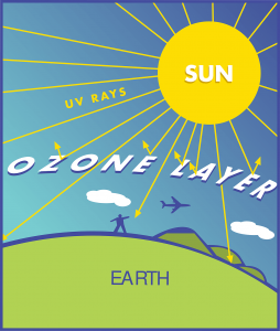
Ozone in the upper atmosphere is our friend.   This thin layer of gas floating between 6 and 31 miles above the earth’s surface protects life on our planet by filtering harmful ultra violet rays from the sun.

Ground level ozone is another story.  This insidious byproduct of automobile exhaust can damage living tissue just as it’s relative in the upper atmosphere protects it. Even the chemical structure of ozone looks like it would be friendly enough: it is just like oxygen, O2, with one more oxygen atom added to make O3 -a super oxygen!

But as vital as oxygen is to sustain human life, too much can be deadly.  To understand why, think about the effect of oxygen on a forest fire on a windy day or what happens to an unpainted iron fence -even in Arizona where it rarely rains.

Rusty BoatWhen wood burns or iron rusts, oxygen is at work in a process called oxidation. Oxidation can turn a battle ship into a heap of rust and kill all the bacteria and algae in your swimming pool.

In fact our immune system uses the deadly effects of oxidation to fight off disease.  Our white blood cells release powerful oxidizing chemicals like hydrogen peroxide and what are called, “reactive oxygen species,” or ROS, that kill and even digest invading pathogens.  This is great when you need to get rid of an infection, but we don’t want these chemicals loose in our bodies digesting us.090114_2215

To keep our white blood cells from eating holes in our lungs and liver, anti-oxidants are produced that are capable of neutralizing the oxidizers and preventing damage. Important antioxidants include vitamin A, vitamin E, beta-carotene, vitamin C, and glutathione as well as many other compounds found in food, especially vegetables and fruits.

When chronic inflammation occurs as a result of injury, infection, allergies, or immunologic processes, excessive amounts of these oxidizing chemicals are produced creating a condition called “oxidative stress”.  This stress can contribute to the pathogenesis of a wide variety of disease states including heart failure, atherosclerosis, and cancer as well as to the normal process of aging.

The role of diet and vitamin supplementation in the treatment and prevention of chronic disease is an important subject and one that I will review in more detail in a later post.   For now, the point I would like to make is that oxidative exposure from external sources can overwhelm our anti-oxidant resources and can contribute to the development and exacerbation of chronic disease.

Which brings us back to ozone.

Ozone is a killer oxidizing agent – powerful enough to be used commercially to sterilize water supplies. It is definitely not something you want to spend much time inhaling.  Exposure above as little as 100 ppb (parts per billion) can be harmful, causing symptoms such as nose, eye, and throat irritation, coughing, wheezing, shortness of breath, painful breathing, nausea and headache, and has been linked to increased incidence of asthma, bronchitis and heart disease.  Long-term exposure has been linked to increased risk of death from lung disease.

Untitled design (36) (1)According to the Maricopa Air Quality Department, ozone levels in our area are can reach unhealthy levels on “hot, sunny days when there is little wind”, which pretty much describes most days in Phoenix from May until October. For example, about a week out of the month of July, 2013 were under a high pollution advisory and health watch for ozone. This year the American Lung Association ranked Phoenix as the 11th most polluted city in the US for ozone.

In recognition of the adverse health effects of ozone, air quality guidelines have been established by the World Health Organization, European Union, and the US Environmental Protection Agency (EPA).   In 2010, the EPA announced proposed revisions to the National Ambient Air Quality Standard (NAAQS) for ozone with the following statement:

EPA proposes that the level of the 8-hour primary standard, which was set at 0.075 μmol/mol in the 2008 final rule, should instead be set at a lower level within the range of 0.060 to 0.070 μmol/mol, to provide increased protection for children and other ‘‘at risk’’ populations against an array of ozone – related adverse health effects that range from decreased lung function and increased respiratory symptoms to serious indicators of respiratory morbidity including emergency department visits and hospital admissions for respiratory causes, and possibly cardiovascular-related morbidity as well as total non- accidental and cardiopulmonary mortality…

In addition, the Air Quality Index (AQI) was developed by the EPA to explain air pollution levels to the public. Using this scale, eight-hour average ozone levels of 85 to 104 nmol/mol are considered “unhealthy for sensitive groups,” 105 nmol/mol to 124 nmol/mol as “unhealthy,” and 125 nmol/mol to 404 nmol/mol as “very unhealthy.. The current AQI for Maricopa county and surrounding areas can be found at

http://alert.fcd.maricopa.gov/alert/Google/v3/air.html

Ozone exposure can have a significant negative impact on lung function, particularly in athletes involved in outdoor sports, a topic I will explore further in the next post.

Exercise Induced Bronchospasm: Treatment

As every athlete involved in an aerobically taxing sport knows, effective breathing can be key.  Muscles will not keep working  (and you won’t keep going) if you can’t breath. Oxygen delivery to working muscles depends on several factors, but the ability to rapidly move a large volume of air in and out of the lungs is critical.   When oxygen demand exceeds supply, an effort can continue for only a short period before you must slow down or stop.   No gas, no go. That’s the law!

Exercise induced bronchospasm (EIB) causes tightening of involuntary muscles surrounding medium to small airways.  This narrowing of thousands of tiny airways limits the rate at which air can be moved into and out the lung.  The “button” that turns on brochospasm in susceptible athletes is a receptor in the lining of the airways that respond to rapid changes in the temperature and humidity of the airway, conditions that frequently occur with such sports as running and biking out of doors.   Rapidly breathing cold, dry air is a particularly strong trigger.  Fortunately, the airways not only have an “on” button that when pushed tells the airway muscles to tighten, but also an “off” button that will relax the tightened airways, relieving the obstruction and allowing air to flow freely.  This muscle- relaxing button is called a beta-receptor.   Not only are these beta-receptors found throughout the airways, but also in the heart and nervous system where, as you might imagine, they have different effects.

The most important medication used in the treatment of asthma is albuterol, a beta agonist.  In other words albuterol “pushes” beta-receptor buttons causing rapid relaxation of the airway muscles and bronchodilation.  Because of the beta-receptors in the heart and nervous system, albuterol can also cause an increase in heart rate and a sensation of nervousness.

Not surprisingly, the most frequently used treatments for EIB is albuterol.   Two inhalations of albuterol fifteen minutes prior to an exercise cession will provide prevent bronchospasm for up to four hours.   Albuterol can also be used when needed to provide rapid relief of asthma symptoms such as wheezing, chest tightness, and shortness of breath within five minutes.   Albuterol is so effective in preventing exercise-induced bronchospasm that response to treatment with albuterol is often used as a test to confirm EIB.   If a patient is suspected of having EIB but does not improve with albuterol before exercise, an alternative diagnosis should be considered.

Albuterol is very effective in preventing symptoms of EIB but it does have limitations.  In athletes who train daily, albuterol can become less effective over time so that, not only does it become less effective in preventing EIB but can also become less effective during an asthma attack.  This is unlikely to be a problem if albuterol is not used daily.

Because of the concerns associated with daily use of albuterol, it is suggested that an athlete with EIB who exercises daily use an inhaled corticosteroid in addition to the albuterol.  Although steroids are not bronchodilators, they are very effective controllers of inflammation and are felt to maintain the effectiveness of albuterol after several weeks of daily use; inhaled steroids may also effectively control EIB without the need for albuterol.

Another medication that has been used for EIB is monteleukast or Singulair.   Monteleukast is preferred by many because it is a tablet (chewable for children) rather than an inhaler, can be used daily, and does not have the concerns associated with an inhaled steroid.  It does not benefit all who use it and many find it significantly less effective than albuterol or an inhaled steroid.

Albuterol is called a short-acting-beta agonist (SABA) because of it relatively short (four-six hours) duration of action.  Long-acting-beta agonists are also available which provide protection from EIB for up to 10 hours.   This would seem to be ideal for many athletes although because of the concern about loss of effectiveness with daily use and the possibility that this kind of medication could mask a worsening asthma attack, it has been recommended that LABAs not be used without the concomitant use of an inhaled corticosteroid.  Fortunately, there are several products available that combine an inhaled corticosteroid with a LABD including Advair, Serevent, and Dulera.   The LABD in Serevent and Dulera can work as quickly as albuterol and can therefore be used as a very effective daily treatment for EIB.

Several non-pharmacologic treatment options may be effective in some with EIB. These include warming up slowly before a hard workout to create a “refractory” state in the airways, preventing bronchospasm and wearing a mask to limit exposure to cold, dry air.

For patients with allergies who also have EIB, adding an antihistamine may be helpful.

Exercise Induced Bronchoconstriction: Diagnosis

On the surface, making a diagnosis of exercise-induced bronchoconstriction (EIB) should not to be too hard. After all, having an asthma attack while exercising would make the diagnosis fairly apparent.  However, as I mentioned earlier, self reported symptoms associated with exercise such as shortness of breath and chest tightness can be associated with a number of causes, both respiratory and non-respiratory, including just being out of shape.   In fact, in one study, approximately half of elite athletes with symptoms suggestive of EIB were found to have normal lung function.  Even more interesting is the fact that nearly half of the athletes who did not feel that they had EIB in fact tested positive for it.

Because symptoms alone are not sufficient, an objective test is required to make an accurate diagnosis.  The best tests for EIB either directly measure airway changes during exercise or produce the inflammatory changes in the airways that occur in patients with EIB and therefore serve as a surrogate for an exercise challenge.

An exercise challenge starts with a baseline pulmonary function determination with serial lung function measurements taken following a period of prescribed exercise.   A typical protocol might include running on a treadmill for 6-8 min at 80-95% of an athlete’s calculated max heart rate.  This level of exercise requires significant effort but is required to produce changes associated with EIB in highly trained athletes.  Following the effort, lung function values are recorded at 5, 10, 15, and 30 minutes and compared with the pre-challenge numbers.  A 10% or greater drop in lung function persisting for at least 15 minutes after exercise has stopped is diagnostic of EIB.

Because EIB is triggered in some athletes by inhalation of cold, dry air, a weakness of an exercise challenge on an indoor treadmill is indoor humidity that may be significantly different form the sport environment.  For this reason, sport and environment specific challenges (field challenges) have been found to be more sensitive than tests run in controlled indoor environments.  This is particularly true for those involved in winter sports such as ice skating and skiing and may very well be important for athletes who train in a desert environment such as Phoenix.   In some, the level of intense exertion present in competition may not be reproduced exercising in a calm, indoor setting, producing falsely negative results.

Several alternatives to an exercise challenge have been developed including what is called Eucapnic Voluntary Hyperpnoea or EVH.  In this test an athlete breathes a specially formulated, dry, gas mixture at a rapid rate to replicate the conditions of a hard exercise cession.  This is currently the only test recommended by the International Olympic Commission to identify EIB in Olympic athletes.  Unfortunately, the test requires specialized equipment and is not without some risk of precipitating a severe asthma attack.  For this reason the test is not widely available, in spite of the IOC recommendations.

Although an objective diagnostic test for EIB is recommended when possible, another approach to is to treat the athlete with an asthma regimen and see if symptoms improve. I’ll discuss more about this in the next post when I review treatment of EIB.

Exercise Induced Bronchoconstriction: What, When, and Where

Exercise-induced bronchoconstriction or bronchospasm (EIB) is defined as acute airway narrowing occurring as a result of exercise.  In the previous post I discussed that exercise, particularly vigorous, aerobic exercise, frequently triggers symptoms in patients who have a diagnosis of asthma but can also cause asthma symptoms, wheezing, shortness of breath, cough, and chest tightness, in athletes who have never had asthma.

Feeling very short of breath after a hard aerobic workout does not mean that you have asthma.  More often it means you are a bit out of shape.  So how can we tell the difference between symptoms caused by going too hard and symptoms caused by EIB?

One difference is that symptoms of EIB do not track with heart rate.  Getting out of breath because of de-conditioning or exercising beyond your aerobic capacity occurs when you are not able to deliver enough oxygen to your muscles to meet their demand.   Your heart beats rapidly and you breath faster and deeper to try to deliver more oxygen to meet the demand. But once you reach your limit, oxygen-starved muscles cannot keep going and you have to slow down or stop.  When you do, your heart rate and breathing slows and the shortness of breath and sensation of air hunger quickly improves.

With EIB however, symptoms do not usually begin until well into an exercise cession, and most importantly, will continue for 30 to 60 minutes after exercising has stopped.

In fact, it is believed that rapid breathing during exercise has a cooling and drying effect on the lining of the airways, which in patients with EIB triggers inflammation.  This inflammation is similar to an allergic asthma attack and will continue after exercising has stopped.  Because cooling and drying of the airways triggers inflammation, symptoms of EIB are more likely to occur during periods of exercise out of doors when the air is cold and dry.   Other environmental conditions that have been found to contribute to EIB include high ozone and particulate levels in the air.

Cold, dry air with high ozone and particulate levels describe conditions frequently encountered in Phoenix and other desert communities in the fall and winter. Athletes involved in aerobic sports requiring high respiratory volumes over an extended period of time such as runners and cyclists are most vulnerable. In fact, exercise induced bronchoconstriction occurs in up to 15% of distance runners.

I will review diagnosis and treatment options for EIB in future posts.

Exercise Induced Bronchoconstriction: Asthma by Any Other Name

Recently, the American Thoracic Society published new clinical practice guidelines for exercise-induced asthma. This is an important subject, particularly for children and adults who are involved in sports or who exercise regularly (which should, of course, be everyone), and so I will review parts of the guidelines over the next few posts.

One of the documents chief recommendations is that the term “exercise induced asthma” be done away with and replaced with “exercise induced bronchoconstriction or bronchospasm (EIB)”.   This recommendation is based on the observation that, although exercise is one of the most common triggers for bronchial narrowing in asthmatics, it also occurs in some athletes (particularly those of the “elite” variety) who have never been diagnosed with or treated for asthma.  To use the term “asthma” may therefore be not only inaccurate but also, possible, unfairly stigmatizing.  By eliminating the term “asthma” and replacing it with “bronchoconstriction”, the diagnosis can be applied to both asthmatics and non-asthmatics alike.

This symantical nuancing highlights limitations in our current understanding of asthma. It is generally agreed that asthma should be considered a condition characterized by chronic inflammation in the airways.  This inflammation is responsible for the phenomenon of airway hyperreactivity, a heightened sensitivity to a variety of environmental triggers including respiratory infections, cigarette smoke, dust, and exercise, which cause reflex tightening of muscles surrounding the airways or bronchospasm.  Bronchospasm produces narrowing of the airways and many of the symptoms characteristic of asthma including shortness of breath, wheezing, cough, and sensation of chest tightness.

Also important in the definition of asthma is reversibility.  Although the narrowing of the airways from bronchospasm can be severe and even life threatening, it is not permanent, and with proper treatment, the limitations and symptoms associated with an asthma attack can be reversed and lung function will return to normal.  In addition, asthma can be a significant problem in a child but remit for a number of years with normal lung functions and only occasional, mild symptoms occurring as a teenager. Under these circumstances, it can be a challenge to answer the question, “do I still have asthma?”

An athlete with EIB has all the characteristics of asthma with the exception of chronicity.   When the athlete is not exercising, lung function is normal.   However, other tests that are used to diagnose asthma may be just as abnormal as in patients with a diagnosis of asthma.

Because many patients who have had a diagnosis of asthma in the past and who now have infrequent symptoms usually have normal lung function, there may be no measurable difference between a patient with mild asthma and a patient with EIB.  And yet under the new American Thoracic Society recommended terminology, EIB is not “asthma”, except when it is.

How to Use an Asthma Inhaler and Nebulizer

The following instructional videos will help you learn the proper use of asthma inhalers, a spacer, and nebulizer

How to Use the Nebulizer Machine – Demonstration

How to Use a Metered Dose Inhaler – Demonstration

How to Use a Diskus for Asthma Relief – Demonstration

How to Use the Pulmicort Flexhaler

How to Use an Inhaler with a Spacer ACP Foundation

Asthma Inhaler at 11 months old, Delaney can do it!!!

Arizona Cough

Cough is one of the most common symptoms prompting patients to see a doctor in the United States with an estimated 30 million trips to the doctor for this problem each year. More than 40% of the patients seen in our allergy and pulmonary practice between November and February complain of cough.

Cough is classified as acute, sub acute or chronic depending on how long the symptom has been present.   Acute cough lasts for less than three weeks and is most commonly the result of an acute respiratory tract infection. Other more serious causes of acute cough include pneumonia and in our clinic in Arizona, coccidiomycosis infection or valley fever.

A cough associated with typical cold symptoms may be called bronchitis, particularly when symptoms last for more than a week. Acute bronchitis is most often caused by a viral infection although other respiratory infections besides viruses, including Mycoplasma pneumoniae, Chlamydophila pneumoniae, and Bordetella pertussis may be involved.  Although most viral infections cause symptoms lasting less than 2-3 weeks, some patients with viral or other upper respiratory tract infections will continue to cough for more than eight weeks after the acute infection.  This persistent cough may be the result of a type of airway injury.  Although the source of the infection is gone, the injury remains and takes time to heal.

Another important cause of acute cough in children in adults is pertussis (whooping cough).  Pertussis is a very contagious disease caused by the bacteria Bordetella pertussis. Before the advent of vaccinations in the 1940s, pertussis was a major cause of severe illness and death among infants and children.  Although cases of pertussis decreased by more than 99% after the introduction of pertussis vaccine, it remains a cause for concern, in part because of the incomplete protection provided by the vaccine and the increasing numbers of children that are never vaccinated.  In fact, pertussis is the only vaccine-preventable disease that is associated with increasing deaths in the United States.  In 2010, more than nine thousand cases of whooping cough were reported in California. At least ten infants died from the infection prompting the health authorities to declare a pertusis epidemic.

Pertusis infection usually begins with symptoms similar to the common cold although after several weeks, frequent and often violent coughing begins. The illness is most severe in infants and young children, particularly in those that have not been immunized. In adults, the only symptoms may be a persistent cough.

In a recent study published in The Journal of Allergy and Clinical Immunology (JACI), the risk of adults and children with asthma developing whooping cough was 1.7 times higher than those without asthma, suggesting that asthma significantly increased risk for whooping cough.

A cough lasting more than 4-6 weeks without a clear history of acute respiratory infection is considered chronic and is most likely the result of one of three conditions: asthma, rhinitis/sinusitis and gastroesophageal reflux disease.

Asthma and rhinitis/sinusitis are frequently the result of allergies and so a history of allergies or a positive allergy evaluation strengthens the likelihood that one of these conditions is behind the cough.

Gastroesophageal reflux disease (GERD) and laryngopharyngeal reflux disease (LPR) are conditions associated with the leakage of stomach contents into the esophagus.  In GERD, stomach acid refluxes into the lower esophagus causing irritation and damage.  Exposed nerves in the esophagus can cause cough as well as pain (heartburn).  In LPR, stomach contents may reach to the top of the esophagus causing direct irritation of the throat and possible sinuses.  The throat and upper airway are lined with cells that produce mucous as well as cells that have hair-like projections or cilia that sweeps the mucous to the back of the throat where it is swallowed.  Acid and protein-destroying enzymes in the refluxed stomach contents inflame and  damage the hair cells, hindering the ability to clear mucous.  The result is pooling of mucous in the back of the throat and recurrent cough to clear it.  It is estimated that 50% of patients with LPR have no other symptom of their condition other than cough and is therefore frequently missed.   GERD and LPR should be suspected if an evaluation for allergies, asthma, and sinus disease is negative and the cough fails to respond to conventional treatment.

The Asthma – Tylenol Link

I have been seeing a number of articles in the news recently reporting a theory that acetaminophen (Tylenol) use in children is linked to the development of asthma.  Proponents of the theory site several lines of evidence.  One is the observation that about 30 years ago parents began to give children with fever acetaminophen in place of Aspirin because of a link between Reye’s syndrome and Aspirin.  This occurred about the same time  that researches began to document a significant increase in asthma cases.  Also, studies have shown that parents of children with a diagnosis of asthma report given their children acetaminophen more frequently.

There are problems with using these observations to conclude that acetaminophen use causes asthma.  Most importantly is the fact that viral respiratory infections, like the rhinovirus that causes the common cold,  are by far the most important triggers of wheezing episodes in children. Many children with wheezing episodes associated with colds go on to develop true asthma but many do not.  In addition, researchers have suggested that certain viral respiratory infections such as the respiratory syncytial virus (RSV) may produce a type of airway injury that leads to the development of asthma.   Since children that have more frequent colds and associated conditions such as ear and sinus infections would be given acetaminophen more often for pain and fever, it is difficult to determine the true link.  In other words, is the observed increase in asthma the result of acetaminophen use or a change in the frequency and types of infections (as well as, perhaps, expectations of parents) that the acetaminophen is used to treat?   Other considerations include the use of antibiotics and vaccinations, declining family size, urbanisation, and pasteurization: all dramatically changed in the past thirty years.

One point is clear however: Reporting a link between Tylenol use and asthma will attract more readers than noting that sick children are more likely to have symptoms.